First, the brain is organized into functionally specific areas, and second, neurons in different parts of the vertebrate nervous system, indeed in all nervous systems, are quite similar.
Small comparison with Computers
A gross observation between computer’s transistors and human neurons is that there a big difference of numbers:
- trillions of transistors vs billions of neurons.
- 6 orders of magnitude frequency difference (Ghz versus 1kHz for neurons).
- Many many neural types and different types of connections.
- And the digital vs analog and chemical modes of communication.
- Parallel processor abilities.
- Fixed vs plastic architectures But this is comparing with transistors with one higher level object, so this comparison might not be completely fair. They are very different from this point of view.
And only some brain areas are similar to real neural networks.
David Marr’s Tri-level approach
We don’t follow now this approach, a clean separation of the levels is not exactly clear. There are some regions that we don’t know what they are doing at all. Internal state with goal is important, most of the computer systems do not have this internal driver.
See original paper.
This revolution stemmed from physiological work and makes us realizethat the activity of each single neuron may play a significant role in perception,
The Tri-level
Computational Level: what kinds of computation is the system doing? Why does it do these things (highest level things) (e.g. intentions and beliefs with ToM).
Algorithmic Representational Level: how does the system do what it does? What processes does it effectively manipulate?
Physical level: how does the system implement it on the physical level (chemical or physical processes of brain firing).
For the brain, the first level it is debatable (not clear what the brain is trying to solve), and the representational level is just incomprehensible. Neuroscientists usually just measure at the physical/hardware level.
What does the Brain do?
This attempts to answer the computational level problem: Some debated things are:
- Brain creates a model of the environment.
- Having models of the world is useful to predict what they are likely to do and plan accordingly.
- The brain makes internal decisions on different time scales (goals).
- This internal decisions (intents) are used to make actions.
- We want to predict the consequence of our actions on the environment.
- Has intentions or goals and acts on it.
- It is able to evaluate their actions (perhaps survival, safety, or other details) and update the internal model of the world.
- Predictive models are useful, to plan and act on this information (example is gazelle chase for predators)
And we would ask how the brain does all this.
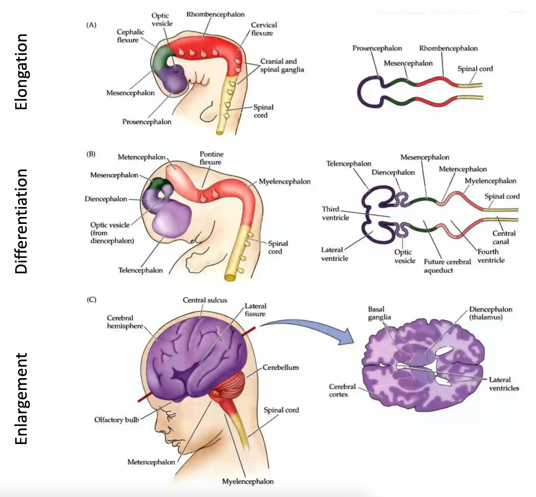
The Development of the Brain
It first starts with an elongated neural tube (this is the precursor. The different parts of development are named by the stage of development, also related to the evolution steps. It is very similar across vertebrates. There are three different development states after the formation of the neural tube.
- Very long tube that then differentiates and becomes bigger and bigger. Diencephalon enlarges. They all form with the same similar process.
Parts of the brain
Most of what we know about the cortex has been discovered by injuries on those parts of the brain. Now we now can use reversible optogenetics to check some parts of the brain precisely: activation/inhibition and recording of brain circuits.
We can now use electrodes in the brain: Deep Brain Stimulation (DBS)(Parkinson’s, Depression, OCD, Epilepsy, …), or subcutaneus parts, or transcranical stimulation bymagnetic/electric fields or ultrasound waves, that can excite some brain areas (not very specific, but not invasive).
Cortex
We have the largest density of cells in the cortex, this is the part of the brain mostly correlated with intelligence. many many abilities correlate with the size of the cortex. Differences between primates is mostly in the cortex.

Image from the slides
Structure of the Cortex
Cortex has very similar structure (also for other mammalian species!) This lead to cortical column theory, parts of the cortex have some specific functional properties. The experiment on cats provides good evidence on this theory. Another good experiment is the one in 1990 by Sur et Al. (Universal swappable computing engines). They proved the plasticity and reorganization capabilities of brain regions during development.
 #### Cortical Theory
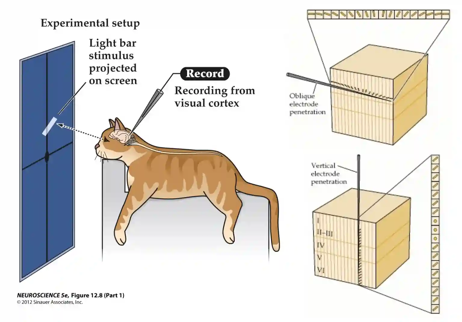
One column is about 0.5mm in diameter and in V1 layers you usually just have dentritic extension by the pyramidal layers in the layers below
Modulating Brain Areas
Neuron-Specific Drug Delivery via Focused Ultrasound Uncaging
- Principle: drugs are packaged in carriers (e.g., nanoparticles, liposomes) that remain inactive (“caged”).
- Focused ultrasound breaks or heats carriers at target sites → local drug release only where ultrasound is applied.
- Advantages:
- High spatial precision.
- Reduces systemic side effects.
- Can deliver neuromodulators, neurotransmitters, or other neuron-specific agents.
Deep Brain Simulation
- Invasive: electrodes surgically implanted into specific brain nuclei.
- Provides continuous or patterned electrical stimulation.
- Clinical uses:
- Parkinson’s disease (reduce tremors, motor symptoms).
- Depression (treatment-resistant).
- OCD.
- Epilepsy.
- Advantage: highly effective in movement disorders.
- Limitation: requires neurosurgery, risk of infection, electrode misplacement.
Transcranial Stimulation
- Uses magnetic, electric, or ultrasound fields applied through the skull.
- Techniques:
- TMS (Transcranial Magnetic Stimulation) → magnetic pulses induce currents in brain tissue.
- tDCS / tACS (Transcranial Direct/Alternating Current Stimulation) → low current applied via scalp electrodes.
- tFUS (Transcranial Focused Ultrasound Stimulation) → acoustic waves target deep brain regions with high spatial precision.
- Applications: Modulate cortical excitability, study brain–behavior relationships, potential therapy for depression, chronic pain, stroke recovery.
Actor Critic Model of the brain
This model that we present here is connected to the theory of how humans integrate the information, explained in Conditioning Theory. We studied actor critic methods in RL Function Approximation. It seems that the brain implements similar modules:

Image from the slides.
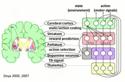
Something very similar to this model happens in the dopamine neurons (providing rewards gives difference in expectations and actual rewards, this is called reward prediction errors.) When we have conditioned stimulus the surge in dopamine happens during the cue, not the actual reward itself. This works for positive errors (not expecting anything while receiving food, where we have a surge of dopamine), and negative errors (not receiving food even if we are expecting it). We analyze reward models in Conditioning Theory.
Distinguishing Cortical Functions
There are mainly three ways that we report to distinguish cortical function.
Functional Evidence
They observed that same cortex was responding in the same way, but different columns in different way, this was functional evidence of those differences.
- fMRI, and responses due to external simulation.
- To manipulations (strokes, and other damages to brain areas, or just cooling).
Functional Organization
The types of cells are different, so the cortices are specialized in this manner, but are able to adapt if their input is different (remember the example of the replaced auditory cortex for visual cortex.) The other is called electrical stimulation mapping.
For example:
- Speech processing (Wernicke) is close to speech production (Broca)
- Speech production is close to motor area.
- Auditory information should be associated with visual information. Hence auditory cortex is proximal to temporal areas..
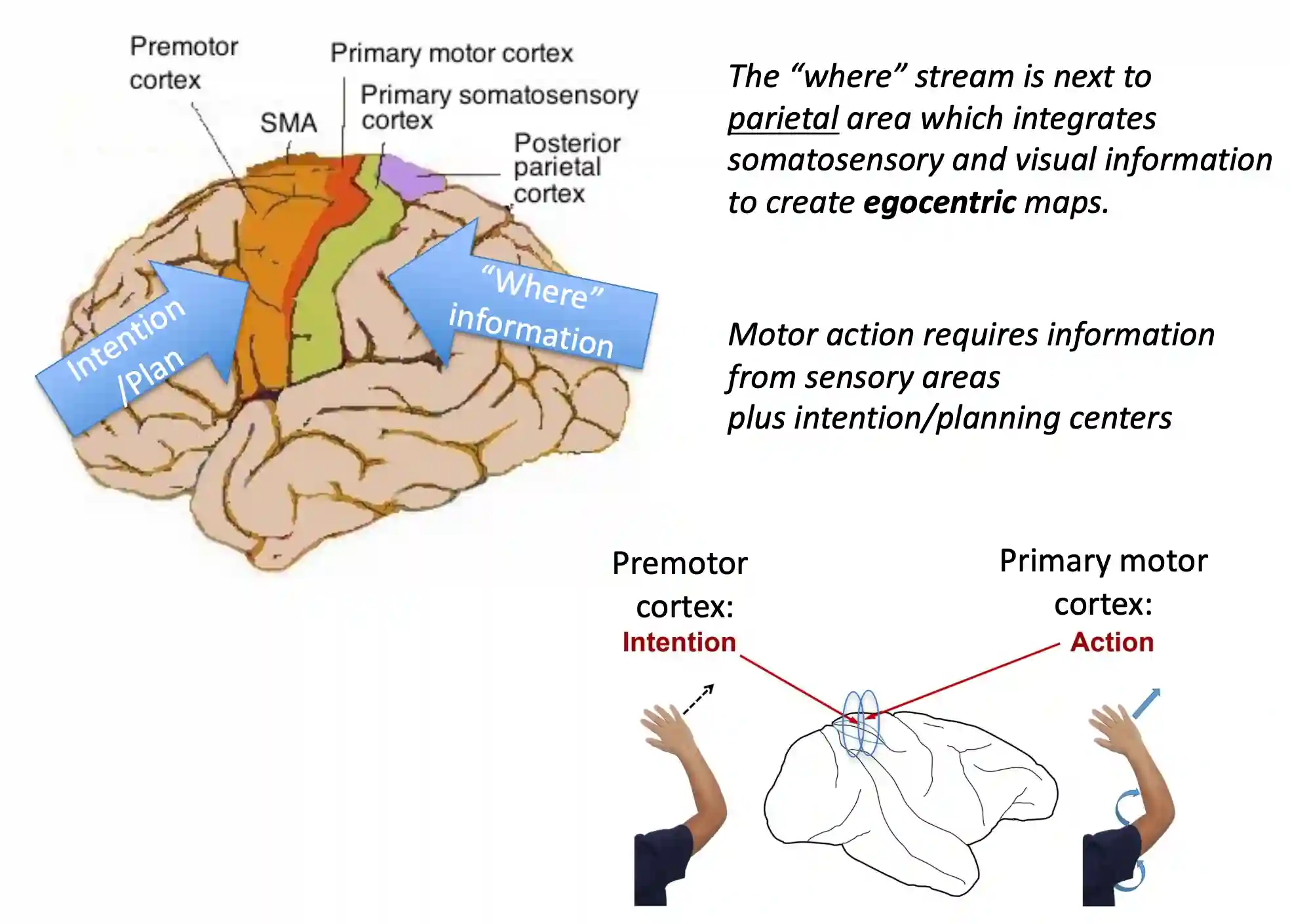
- The “where” stream is next to parietal area which integrates somatosensory and visual information to create egocentric maps. See Human Vision.
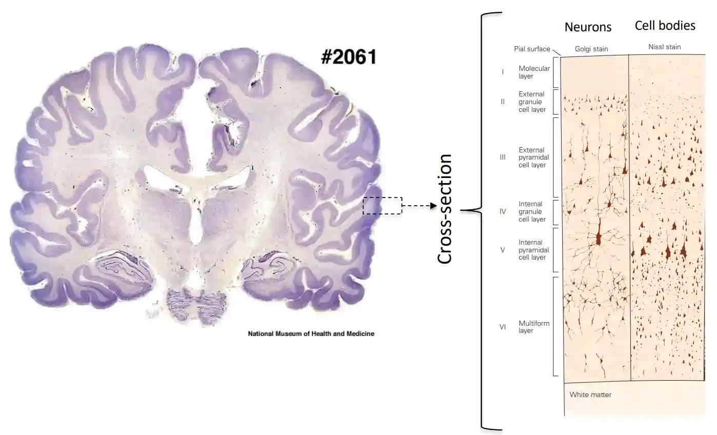
Cytoarchitectonics
Another method to identify brain areas is to see at the cytoarchitectural structure of the cells:
- Pioneered by Korbinian Brodmann → “Brodmann areas.”
- Method: study the size, density, and layering of neurons under a microscope using histological stains (e.g., Nissl stain).
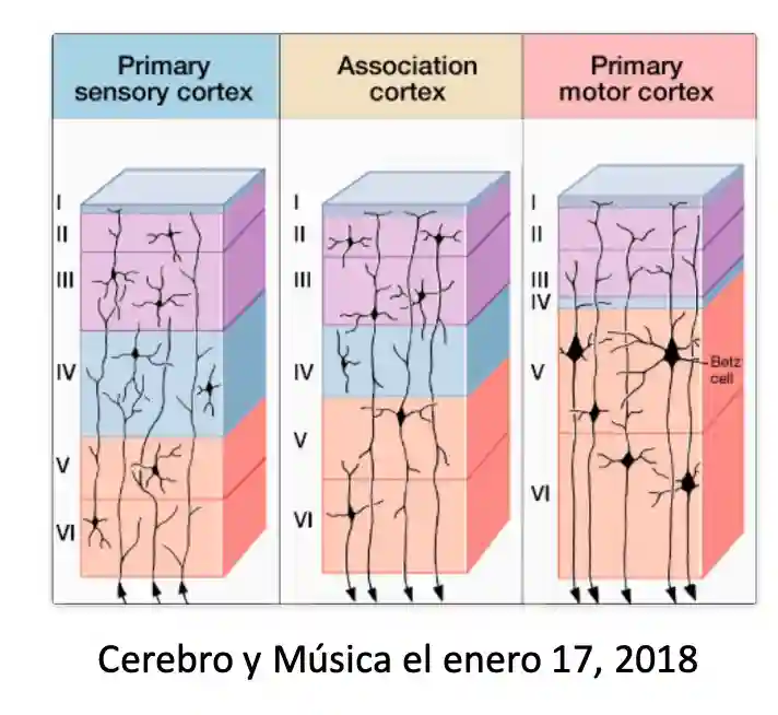
- Contribution: reveals systematic differences in cortical layers (e.g., primary sensory cortex has a thick Layer IV for thalamic input; motor cortex has giant pyramidal cells in Layer V).
- This allows researchers to define distinct structural “maps” of cortex even without knowing function.
There is also a close friend called Myeloarchitectonics (fibers
- Method: stain for myelinated axons (e.g., using Weigert stains).
- Contribution: highlights the density and pattern of intracortical connections.
- Different areas show unique myelination patterns — for instance, primary visual cortex (V1) has a prominent stria of Gennari (a dense band of myelinated fibers in Layer IV).
- This gives an anatomical signature distinguishing one cortical area from another.
Cytoarchitectornics of Motor and Sensory Cortex
For example: Motor cortex:
- The primary motor cortex (M1) is heavily involved in sending commands to the spinal cord and brainstem to drive movement.
- Layer V of M1 is packed with large pyramidal neurons, particularly the Betz cells, which are some of the largest neurons in the brain.
- These neurons project directly to the spinal cord (corticospinal tract), which is why Layer V is so prominent.
Sensory cortex:
- Primary sensory areas, like the primary somatosensory cortex (S1), primarily receive input from the thalamus.
- Thalamic projections mostly terminate in Layer IV, which is why Layer IV is thick in sensory cortices and relatively thin in motor cortex.
- The sensory cortex then processes and distributes this input to other layers for integration and output.
Swappable Computing Engines

Rewiring
-
Creating space in the auditory pathway: They damaged the inferior colliculus, which normally provides the major input to the medial geniculate nucleus (MGN) - the auditory thalamus
-
Forcing visual inputs into auditory circuits: Following neonatal surgical manipulations, a specific population of retinal ganglion cells is induced to innervate the auditory thalamus and provides visual input to cells in auditory cortex
-
Functional Changes: These studies involve making unilateral lesions and inducing retinal inputs into the auditory thalamus (MGN) during early development in ferrets, thereby conferring visual responsiveness on primary auditory cortex (AI)
-
Topographic Organization: The rewired auditory cortex developed visual maps and topographic organization similar to what you’d normally see in visual cortex, though adapted to the constraints of auditory cortical architecture.
These experiments demonstrated that cortical areas aren’t rigidly predetermined for specific sensory modalities. Instead, they showed that the type of sensory input largely determines how a cortical area will organize and function. This challenged the prevailing view that sensory cortices were hardwired for their specific modalities and provided compelling evidence for activity-dependent cortical specification during development.
However:
Cortex has many diverse groups of neurons with different roles and connectivity. There exist major differences in cell types and their connectivity across cortical areas.
Areas of brain processing
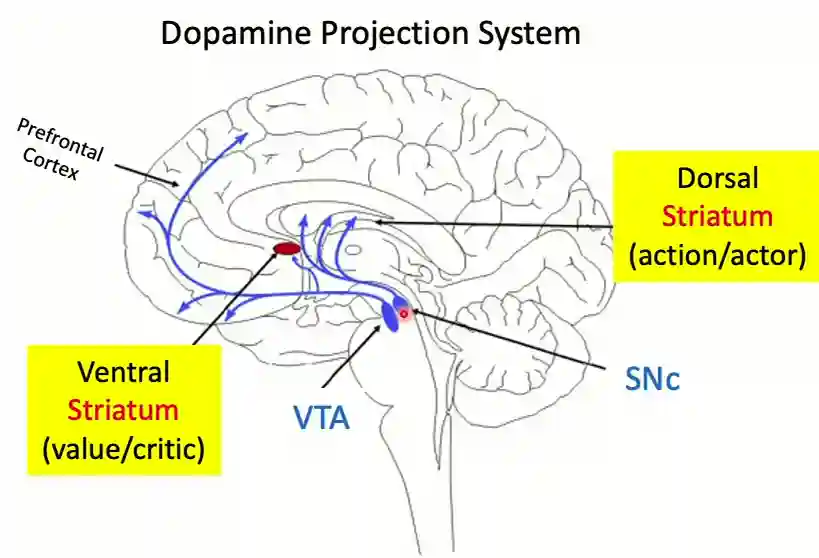
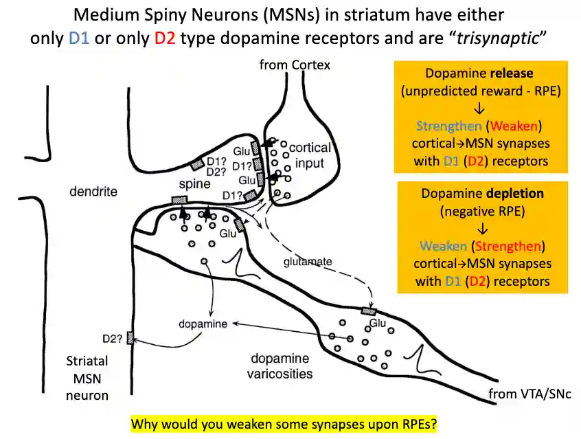
The striatum
The striatum area seems to converge processing from the dopaminergic pathways and the cortical areas. Medium spiny neurons form trisynaptic connections with the cortex and the VTA (ventral tegmental area).
- If the dopamine binds to D1 receptors, it becomes weaker, if D2 the connection becomes stronger. D1 stimulates movement while D2 inhibits it.
- It starts with very complex signal and ends up with a few actuator neurons (some thousands).
The learning loops becomes shorter with time, (From conscious learning to unconscious execution).
There are mainly two pathways in the striatum, the ventral and the dorsal one.
- Ventral: (Nucleus accumbens), is the main region that gets dopamine dignals from the VTA section, this is why we assume this section does reward computation.
- Dorsal: It has controls to the motor and cognitive control parts, so this part decides the actions to take.
And the major inputs to striatum are:
- Cortex (glutamate → excitatory).
- Thalamus (glutamate).
- Dopamine from midbrain (VTA/SNc → modulatory).
 #### Pallidum (Globus Pallidus)
- Output stage of the basal ganglia (striatum zone).
- Constantly inhibits thalamus (a “**brake**” on movement).
- Sends inhibitory (GABAergic) output to the thalamus and brainstem
- When striatum activates the direct pathway → it inhibits pallidum → brake lifted → thalamus excites cortex → action executed.
- When the indirect pathway is dominant → pallidum inhibition persists → action suppressed.
- **Role:** The “final *gatekeeper*” controlling whether cortical action plans actually happen.
#### Pallidum (Globus Pallidus)
- Output stage of the basal ganglia (striatum zone).
- Constantly inhibits thalamus (a “**brake**” on movement).
- Sends inhibitory (GABAergic) output to the thalamus and brainstem
- When striatum activates the direct pathway → it inhibits pallidum → brake lifted → thalamus excites cortex → action executed.
- When the indirect pathway is dominant → pallidum inhibition persists → action suppressed.
- **Role:** The “final *gatekeeper*” controlling whether cortical action plans actually happen.
Very close to the reinforcement learning loop, this is why the actor critic model of the brain makes sense.
- Cortex suggests possible actions based on goals and environment.
- Striatum receives these options and, influenced by dopamine from VTA, learns which are valuable.
- Pallidum implements the “brake system,” allowing only the favored action through.
- The executed action leads to an outcome (reward or not).
- VTA compares outcome vs expectation → updates dopamine signals → reshapes striatal synapses → next time, the system is better tuned.
So pallidum directly filters the correct action. Both are inhibitory, but:
- Pallidum = “which action should happen?” → targets motor thalamus for movement control.
- TRN = “which information should be noticed?” → targets sensory thalamus for attention and gating.
| Feature | Pallidum | TRN |
|---|---|---|
| Main target | Thalamus (DLM / motor thalamus) | Thalamus (sensory and association relay nuclei) |
| Purpose | Motor/action selection | Sensory gating, attention, oscillatory coordination |
| Inputs | Striatum / basal ganglia | Cortex and thalamus |
| Function | Decides which motor commands proceed | Shapes and filters thalamic output to cortex |
| Effect of inhibition | Disinhibits cortex indirectly | Temporarily suppresses thalamic relay to cortex |
The brain stem

Image from Kandel
Medulla
The medulla, the most caudal portion of the brain stem, is a direct extension of the spinal cord and resembles the spinal cord both in organization and function. Neuronal groups in the medulla participate in regulating blood pressure and respiration. The medulla also contains neuronal groups that are early components of pathways that mediate taste, hearing, and maintenance of balance as well as the control of neck and facial muscles.
Pons
The pons lies rostral to the medulla and protrudes from the ventral surface of the brain stem. The ventral portion of the pons contains the pontine nuclei, groups of neurons that relay information about movement and sensation from the cerebral cortex to the cerebellum. The dorsal portion of the pons contains structures involved in respiration, taste, and sleep.
Midbrain
\[...\]The midbrain also contains components of the auditory and visual systems. Finally, several regions of the midbrain give rise to pathways that are connected to the extraocular muscles that control eye movements.
The Thalamus
This is one of the most complex brain areas. It has a role in consciousness, different firing patterns here if you are asleep or not. We can see this section as the routing board of the brain (almost every sense of the human brain passes through the thalamus, but not the sense of smell).
It receives information from sensory areas, but above all from cortical areas. This is also why we think the thalamus plays an important role with regards to consciousness.
The entire cortex is connected to the thalamus, it also controls the communication between different brain regions, between one cortical area and the other cortical area. We have lots of back connections, probably because of attention selection and similar things.
 > Thalamic DBS can rouse patients from minimally conscious state
Thalamic Reticular Nucleus
Here we have lots of GABAenergic neurons that project back to the thalamus, which is why it is believed to act as a gate.
The thalamic reticular nucleus acts as a gateway between thalamus and cortical connections, they are called attention centers (like amygdala) and modulate the thalamic information to the cortex, and most of the connections here are inhibitory by nature.
- Gatekeeper of thalamic relay:
- TRN modulates the flow of sensory information from thalamus to cortex.
- Can suppress or enhance signals depending on attention, state, or timing.
- Control of attention and sensory filtering:
- By selectively inhibiting thalamic neurons, TRN helps focus cortical processing on relevant stimuli.
- Prevents irrelevant signals from reaching the cortex (“sensory gating”).
- Generating rhythmic activity:
- TRN contributes to thalamocortical oscillations, e.g., sleep spindles during non-REM sleep.
- These oscillations are important for memory consolidation and attentional control.
The Pulvinar
We can view this as a second order thalamic connection. The pulvinar is the largest nucleus of the thalamus in primates, sitting at the very back (posterior thalamus).
Unlike the lateral geniculate nucleus (LGN), which gets input from the retina, the pulvinar gets most of its driving input from cortex (especially visual areas like V1, V2, MT, parietal cortex).
- Then, it sends projections back to multiple cortical visual areas.
- It’s basically a “hub” for visual cortex communication.
Researchers think of the pulvinar as a higher-order thalamic relay that:
- Coordinates cortical areas: For example, making sure motion areas (MT) and shape/scene areas (V4, IT) talk to each other efficiently.
- Regulates attention: Helps filter out distractions and enhance relevant stimuli. If pulvinar is damaged, people have trouble ignoring irrelevant visual clutter.
- Synchronizes rhythms: Acts like a “metronome,” helping distant cortical regions stay in step during visual tasks.
The Hippocampus
It sits near the enthorinal cortex (this sends much input into the hippocampus, mainly in two pathways) Dentate Gyrus or dentate region -> CA3 -> CA1 (the output of the hippocampus, connected to the rest of the brain).
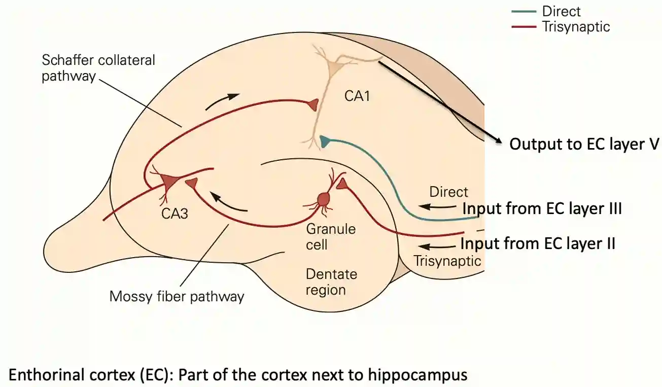
- Entorhinal cortex has about 200k cells (10-20% interneurons)
- Dentate Gyrus has about 1.2kk granule cells. (some sort of a sparse encoder, it is separating patterns)
- 4k basket cells
- 32k Hilar interneurons (20k mossy cells)
- CA3/CA1 about 330k/420k piramidal cells.
- CA3 has many auto-associative recurrent architectures, it is believed to be doing pattern completion after separation with a given context.
The dentate gyrus has much more neurons than other neurons.
Random Functional areas
Mostly the maps to the functions of the brain has been developed by analysis of damages on these regions. A notable example is Phineas Gage’s case on frontal cortex damage. He completely changed his personality. (valuation and planning abilities).
Broca’s area is used for speech production: they can understand sentences but could not produce words.
Wernicke’s area: cannot interpret speeches, they can produce sentences, but they are irrelevant.
Entorhynal Cortex
The researchers performed extensive patch-clamp recordings from over 600 pairs of stellate cells (the primary cell type in MEC layer II that likely includes grid cells, we study those in Memory in Human Brain) in rat brain slices. The main neurons there are stellate cells. Researchers wanted to know: How do these stellate cells connect to each other?
Their main findings were:
1. Absence of Direct Excitation: Despite recording from 644 stellate cell pairs, they found virtually no direct excitatory connections between these cells in mature tissue. Only 4% showed any inhibitory responses to single-cell stimulation.
2. Inhibitory Network Structure: When multiple stellate cells were stimulated simultaneously, 64% of target cells showed inhibitory responses. These were mediated by fast-spiking interneurons rather than direct connections.
3. Developmental Changes: Young animals (postnatal days 10-15) showed rare excitatory connections between stellate cells that disappeared by 4 weeks of age, while inhibitory connectivity matured over the same period.
4. Confirmation with Optogenetics: Using light-activated channelrhodopsin to stimulate populations of stellate cells confirmed that connectivity was predominantly inhibitory.
Object recognition areas
In animals it is possible to use optical techniques to turn on or off specific parts of the neural regions. fMRI uses the activated oxygen levels in certain parts of the human brain, and consumes more energy on those parts. It is a quite precised method. Good for images.
How fMRI works
- Local oxygenated blood has different magnetic properties that can be detected and visualized. It has quite a speed! Some areas are just activated by imagination. This motivated more exploration for understanding functional areas of the brain.
Fusiform Face Area
The fusiform face area was a quite nice discovery: there is a specific region in the brain that activates just by looking at parts of the face.
Parahippocampal Place Area
Or another is parahippocampal place area, that activates for places (houses, landscapes, corners of the room, but doesn’t respond for other things, objects, or abstract art). Probably something that has to do with Place Cells, we studied these in Memory in Human Brain. Identified by Epstein & Kanwisher in 1998 using fMRI. They noticed a region that lights up strongly when subjects look at pictures of places (houses, landscapes, city streets) compared to faces, objects, or scrambled images.
Extrastriate Body Area
Extrastriate body area: responds to body parts (no faces, no random cartoons, yes stickman).
There are other neurons that activate for specific parts of the brain.
Face Neurons
Bruce Desimone and Gross in 1981 identified areas of the brain that activate by faces
Visual Areas
Hierarchical organization of visual processing
This has been the main inspiration for visual processing architectures like Convolutional Neural Network. They start to recognize simple features, and then compose them in more complex shapes. This has been also validated with mechanistic interpretability techniques, and it is overall quite fascinating.
In visual things there are V1, V2, V4 TEO, and cortex areas, in quite organized parts. Mainly in the inferior temporal lobe, which is the “what” pathway for object recognition.
Bidirectional Connections
Convolutional nets are not RNN, they do not use higher contextual information to inform back the small parts. Context is important for humans. Here information is bidirectional, this is the main difference for these biological connections compared to networks like Convolutional Neural Network. The hypothesis is that it helps to interpret what is seen, some feedback signal, somehow makes me think about (Hofstadter 2007). This gives grounds to explain some of the visual illusions.
One hypothesis we have is that it enlarges the receptive field, and uses attention on the part that could be more interesting.
Main roles:
- Attention & prediction: higher areas tell earlier areas what to look for (“expect a face here”).
- Context filling: feedback helps sharpen perception (e.g., blurry letters become readable in context).
Visual information pathways
In visual processing, there are two streams of connections that give different information on
- Where (Dorsal, vestibular information about body positions (eye, arm etc…), that help in action planning)
- They recognize the objects, but not able to know spatial position between them.
- What (Ventral, more in object recognition)
- People that have damage here are not able to recognize the objects, but just know that they exists.
- But they would be able to manipulate the object, not consciously being able to recognize it.
- They share the first layers (v1,v2) for low level details. They tested this in object discrimination and landmark discrimination tasks with dorsal and ventral damages.
We also study the role of learning, since many learning activities pass through the vision part, that is articulated in Memory in Human Brain. If you want a better understanding of vision in general see Human Vision.
Dorsal stream
Looking at the visual stream, we observe that the organization of the parts is not casual, the information gets into the somatosensory area. Posterior parietal cortex uses the information to build model of yourself (egocentric maps), and it is quite close to the cortices, close to the planning part of the brain.
- Premotor area is for physical intention formation
Ventral Stream
The other stream is close to the broca’s area for speech processing and speech interpretation, to associate these two streams. Visual area is heavily linked to the auditory one, but not much with the speech planning area, this explains also why we have these similitudes.

Example of organization of speech areas (auditory and speech information)
Representation of Intentions
Anterior regions represent goals and intentions because they need abstract, context-dependent, temporally extended information that guides posterior motor circuits for execution.
- Premotor and motor cortex (posterior frontal regions):
- Represent specific movements: which muscles contract, sequence of joint actions.
- More anterior regions (prefrontal cortex, especially dorsolateral PFC):
- Represent higher-level plans, goals, and intentions.
- Not tied to a particular movement; more about what you want to achieve.

Goals and intentions in the human brain
References
[1] Hofstadter “I Am a Strange Loop” Basic Books 2007