Synapses are the connections that exist between one neuron and another, so we can think of them as the communication channel between neurons.
Gap Junctions
Electrical based
These are also called Gap Junctions
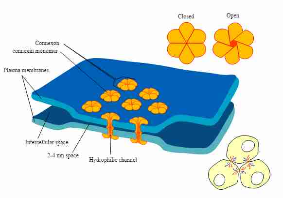
These are more direct connections between neurons, allowing excitation ions to pass through quite directly (this is the difference compared to chemically based ones). It’s a circuit more similar to an electronic one because it’s faster. The end of the presynapsic part is called axon bouton, or axon terminal.
Another characteristic of these kinds of synapses is that they are two-way channels.
They are usually necessary for fast processes, such as when we want neurons to fire together, like in arc reflexes.
Electron-myscroscopy has a definition of 10nm, so it’s quite a challenge to see these gap Junctions of about 3.5nm, between each other..
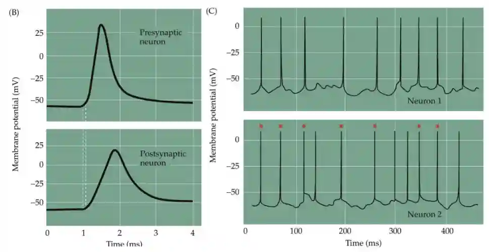
We have almost instantaneous spike transmission, between the neurons (almost synchronized activity). We can interpret this as if it is passive propagation.
Speed of transmission
We can observe that most of the action potentials are transmitted to the other, so they are heavily paired.
Neurotrasmitters
What is Neurotransmitter?
They have four key properties.
-
They are stored in synaptic vesicles at the presynaptic terminal
-
Released in response to calcium influx during action potentials
-
Bind to specific receptors on the target cell.
-
They need to be present in the synaptic clepft.
-
Are rapidly removed from the synaptic cleft (either by reuptake or enzymatic breakdown)
Examples are glutamate, GABA.
E.g. Alcohol and Cocaine are not neurotransmitters as there are no bindings to receptors, but they are called neuromodulators.
Agonist and Antagonist Transmitters
They have antagonists and agonists meaning they make receptors close or open. Two main transmitters are small molecules (glutamate and GABA) and peptide neurotrasmitters. Glutamate is excitatory because its receptors allow positive ions to enter the postsynaptic neuron, making it more positively charged and thus more likely to fire an action potential. Conversely, GABA is inhibitory because its receptors allow negative chloride ions into the neuron, or cause positive ions to flow out, making the neuron more negative and less likely to fire.
Small Molecule Transmitters
They usually have precursors before being released. These are the classic Glutamate GABA etc…
They are small and lightcore (their core is light, instead of dense as peptide transmitters). These are rapidly synthesized on-demand and stored in small synaptic vesicles (40-50 nm diameter) ready for immediate release. They are light 40-50nm vesicles in EM scans.
Neuropeptide transmitters
Chains of amino acids (short proteins) used as chemical messengers in the nervous system. Made in the cell body (soma) — not locally in the axon terminal, this is the main difference compared to other neurotransmitters. Not “point-to-point” like glutamate or GABA → they can diffuse farther and act on a broader range of targets (“volume transmission”). Effects are slower, longer-lasting, and modulatory, and act on the entire system usually. They’re not taken back up by transporters, but broken down GALAMIN is one example.
Recycling of Neurotransmitters
It takes about 10-20 seconds to recycle the neurotransmitter, so it’s quite a slow process. The whole process takes about one minute
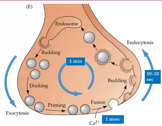
There are many diseases that interfer with this process (for example tetrodotoxin (TTX), which blocks the release of neurotransmitters). They did an experiment with HRP to prove it (so you can see the recycling part).
The advantage of having a recycling cycle (slower for neuropeptides) is that they give some time and spatial scale modulation.
Neurotransmitter based Synapses
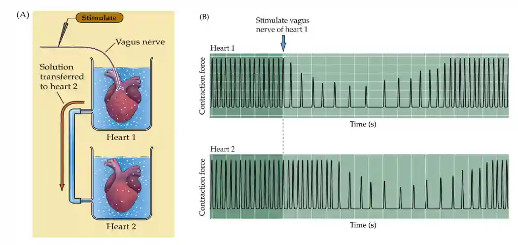
Löwi experiments
Experiment done by Loewi in 1921, where he took two frog hearts and stimulated the first one, then put the liquid in the second one, and it started to beat. It was a groundbreaking study that provided the first direct evidence for chemical neurotransmission Loewi’s work opened the door to discovering the vast array of neurotransmitters we know today and fundamentally changed how we understand brain function, leading to his Nobel Prize in 1936.
This was the first proof of the existence of chemical synapses.
He was studying the heart at the time. He discovered chemical based transmission of synaptical information.
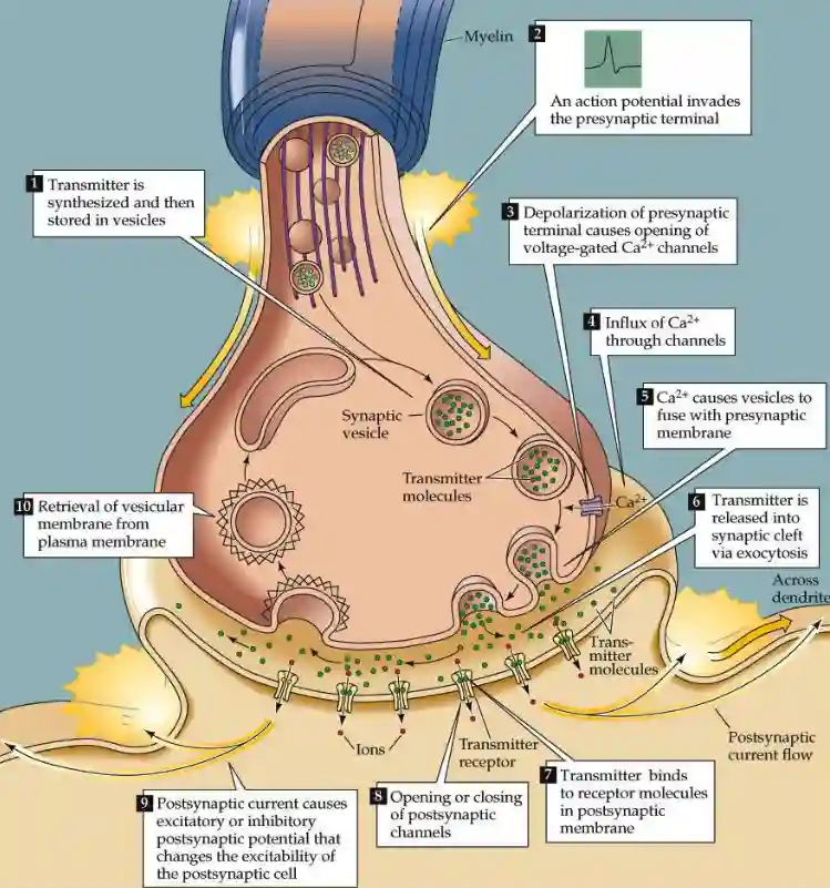
Steps of a Synapse
We open Calcium channels at the pre-synaptic neuron, and then we have the vesicles that contain the neurotransmitter.
This leads to vesicle fusion with the membrane and the release of the neurotransmitter in the synaptic cleft.
Sodium is already used for depolarization (more granular). While Calcium is lower inside the cell, so it is a clear switch, another degree of freedom that is used. Calcium modifies molecular properties of proteins that lead to the fusion of the vesicle.
See that transmitter molecules are recycled with endocitosis.
Chemical-Based
These are slower gates, the classic ones, which we then call neurotransmitters. When there is an action potential, vesicles are released into the intra-cellular space. On the other neuron, there are receptors that pick up these neurotransmitters and activate accordingly.
They are also quite slow diffusion takes about a couple of ms to reach the other side.
The interesting thing about this method is that the receptor can change gates to determine how much it cares about this new information. (So, if it cares about that signal, it can increase the number of gates; otherwise, it can decrease them—at least, that’s the theory.)
When the neuro-trasmittor binds with the post-synaptic neuron, it triggers a release of ions in the other cell that change the membrane conductivity, and thus the voltage (see Firing-rate based Network models).
The cerebral cortex is one of the most important parts of the brain. It consists of 6 uniform layers of neurons that follow a similar pattern.
Layers 1 and 4 receive input from higher cortical areas, layer 4 from sub-cortical areas, and layer 6 from internal structures like the thalamus.
Correlations with Action Potential Timing
Between two communicating neurons, there is a fairly evident correlation that implies an increase in the strength of the connection (synaptic strength) with the timing of the spikes.
We also study this in Memory in Human Brain for potentiation of the Schaffer collatera.
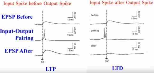
These are called Long-Term Depression and Long-Term Potentiation.
EPSP stands for Excitatory Post-Synaptic Potentiation. In the image, we see that if the second neuron activates after the first neuron has activated, the signal tends to strengthen; otherwise, it weakens.
The weakening makes sense because it’s like I’m giving the signal back to you (even if not necessarily connected), or the other neuron is just tired, lol. We will better explain these phenomena in the following section.
Why calcium
We have learned that calcium is very important for triggering vesicle release of neurotransmitters in the synaptic clepft. This is because under standard situations there is no Calcium in the axon, and this is a clear switch indicator to release transmitters. While for sodium it is difficult to have a clear shift. It is some sort of state change. See Neural mechanisms.