Introduction to the Course
What is a neural system?
A neural system is an intricately organized network of specialized cells—primarily neurons, along with a variety of supportive glial cells—that processes and transmits information via electrical and chemical signals. In biological organisms, such systems underpin the entire nervous system, coordinating functions that range from basic reflexes to the complex interplay of perception, thought, and behavior. Early studies in neurobiology revealed that even simple neural circuits can generate coordinated responses, while modern neuroscience has shown that vast, hierarchically structured networks (such as the central and peripheral nervous systems) are responsible for the rich tapestry of animal behavior and cognition
Types of functions
Sensory systems represent information about the state of the organism and its environment. Motor systems organize and generate actions. Associational systems link the sensory and motor sides of the nervous system.
The Gene look
At the beginning, scientists attempted to look for details of what made humans what we are, what made us intelligent. One clear indicator that influences that is the generic code.
- Worm has around 20k genes.
- Drosophila around 15k
- Mouse 27k
- Humans 35k Most of the genes are expressed in the developing and living brain.
Neither the size of the brain. Another possibility is the ratio between the size of the brain and the body size. Another possible parameter is the density of the neural connections.
In the same way it is not:
- Number of neurons (mouse has like 0.1 billions, humans like 100 billions of neurons, and whales have like 200 billions).
- Heavyness of the brain
- Relation to size That identify how complex is the brain and neural system (ratio is the best indicator though)
The post-genomic era
One of the most promising dividends of sequencing the human genome has been the realization that one or a few genes, when altered (mutated), can begin to explain some aspects of neurological and psychiatric diseases.
Genetic Engineering
For example, viruses can be engineered to transfect a cell or group of cells to express a fluorescent protein of some type. A promoter is a region of DNA that initiates transcription of a particular gene (a promoter is a DNA sequence that turns the gene ‘on’). Promoters are located near the transcription start sites of genes, on the same strand and upstream on the DNA.
A reporter gene (often simply reporter) is a gene that researchers attach to a regulatory sequence of another gene of interest. The reporter is only expressed in those cells that express the gene. Certain genes are chosen as reporters because the characteristics they confer are easily identified and measured, or because they are selectable markers (e.g. such as green fluorescent protein, GFP).
Some history: Reticular Theory vs Neuron Doctrine
The late 19th century witnessed a debate in neuroscience between Camillo Golgi and Santiago Ramón y Cajal, two pioneers whose opposing views shaped our understanding of the nervous system. This debate centered on the structural and functional organization of neurons, culminating in their joint reception of the 1906 Nobel Prize in Physiology or Medicine.
Golgi’s Reticular Theory
Golgi proposed the Reticular Theory based on his staining techniques (see #Staining methods), which held that:
- The nervous system functions as a continuous network rather than being composed of individual cells.
- Neurons are physically fused together into a syncytium, forming a vast, interconnected reticulum.
- Electrical impulses travel freely through this continuous structure, without the need for discrete cellular units.
Golgi’s model was conceptually aligned with other biological systems of the time, such as the circulatory system, which was known to form a continuous network of blood vessels.
Cajal’s Neuron Doctrine
Santiago Ramón y Cajal, a Spanish neuroscientist, refined and expanded upon Golgi’s staining method, using it to produce detailed drawings of neurons. His meticulous observations led him to reject the Reticular Theory in favor of the Neuron Doctrine, which proposed that:
- The nervous system is composed of discrete, individual cells called neurons.
- Neurons communicate through specialized contact points (later identified as synapses by Sherrington) rather than through direct cytoplasmic continuity.
- Information flows in a directional manner from dendrites to axon terminals, supporting the concept of unidirectional signal propagation.
Cajal’s work provided strong anatomical evidence that neurons were separate entities, fundamentally reshaping the understanding of neural communication.
Their conflicting views were most prominently displayed at the 1906 Nobel Prize ceremony, where both gave speeches that reinforced their respective positions.
The structure of the Neuron
Main Structural Parts
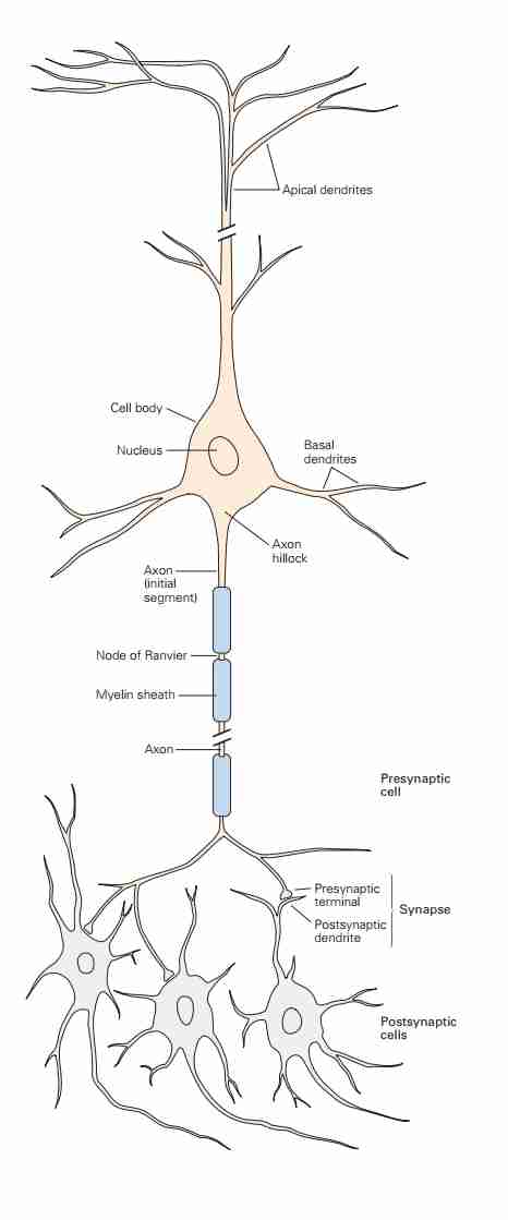
We can identify three main parts regarding the structure of a single neuron:
- Axons, which are responsible for sending activation potential signals externally, communicating with other cells. The signal originating from the axon starts from a section called the initial segment.
- They travel for around few hundred micrometers typically, most of them no more than few millimeters in length.
- A few special cases can be up to a meter long, like the axon that goes from the spinal cord to the foot.
- Dendrites (also called dentritic branches or processes), which are responsible for receiving signals from other neurons.
The number of inputs that a particular neuron receives depends on the complexity of its dendritic arbor: nerve cells that lack dendrites are innervated by (thus, receive electrical signals from) just one or a few other nerve cells, whereas those with increasingly elaborate dendrites are innervated by a commensurately larger number of other neurons.
Which means that the complexity of the input in these cells is related to the branching of the dendrites. These inputs can vary from 1 to 100'000 for each nerve cell. We call the end of an axon the pre-synaptic terminal and the end postsynaptic specialization.
The axons and dendrites are not directly connected; there is a small space between them called the synaptic cleft (the discovery of this was astonishing; in the past, it was thought that the brain was a continuous entity (see above), but instead, we have small discrete units, discovered through the silver staining methods of Golgi, see #Staining methods). The information in this pre- and post-synaptic space is managed by neurotransmitters.
Typically, for a single neuron, we are dealing with dimensions on the order of micrometers.
We can also analyze the neuron from the perspective of its main functional parts, in which case there are four, as illustrated in the image for different types of neurons.
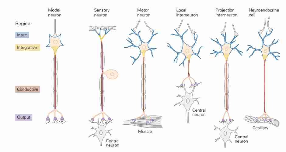 The magically interesting thing is that the connections can be vastly different, yet they ultimately adhere to a fairly coherent concept of the parts (in a sense, we also find here the reasoning by abstraction common in computer science—likely, from the physical perspective of neurotransmitters, there are differences, but they offer the same interface for attachment, so to speak).
Parts of the Soma
- Endoplasmatic reticulum (stores the calcium, which is important for the activation potentials)
- The endoplasmic reticulum is a network of membranous tubules within the cytoplasm of eukaryotic cells, continuous with the nuclear membrane. It usually has ribosomes attached and is involved in protein and lipid synthesis.
- Dentrites: these are the inputs of the cells.
- Golgi apparatus:
- The golgi apparatus is a complex of vesicles and folded membranes within the cytoplasm, it is involved in secretion and intracellular transport.
- Vesicles: (pre and post synaptic parts)
- Axon: thread-like part of the cell that transmits impulses from one cell to another.
- Mitochondria
- Mitochondria are the energy factories of the cells. The energy currency for the work that animals do is the energy-rich molecule adenosine triphosphate (ATP). The ATP is produced in the mitochondria using energy stored in food.
- Ribosome
- A Ribosome is a minute particle consisting of RNA and associated proteins found in large numbers in the cytoplasm of living cells. Ribosomes bind messenger RNA and transfer RNA to synthesize polypeptides and proteins.

From Purves
- A Ribosome is a minute particle consisting of RNA and associated proteins found in large numbers in the cytoplasm of living cells. Ribosomes bind messenger RNA and transfer RNA to synthesize polypeptides and proteins.
See that neurons are separated! The synaptic endings (called terminal boutons) have little spaces, called synaptic clefts where neurostrasmettitors are released. We call it presynaptic and postsynaptic connections.
Classificazioni dei neuroni
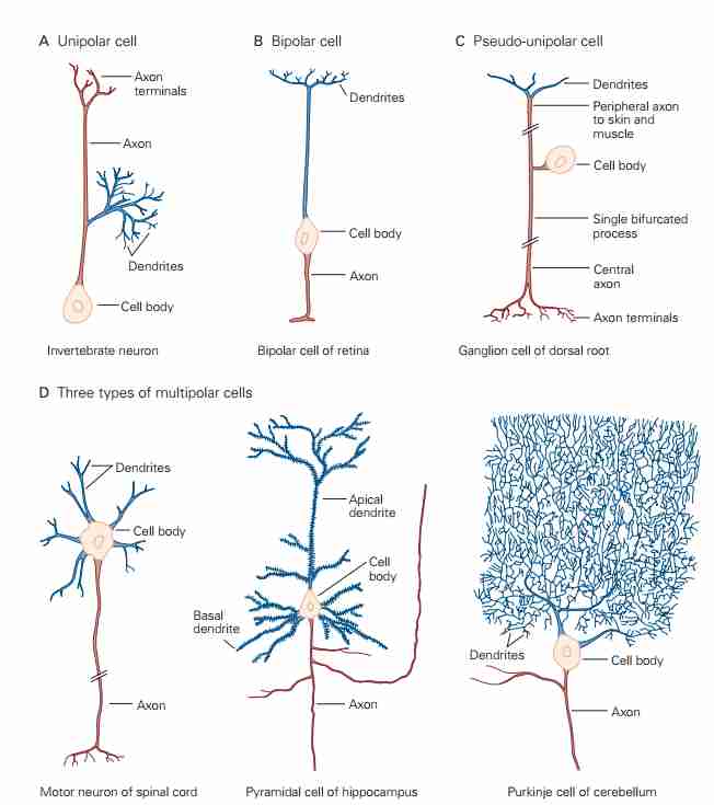
Ci sono molte tipologie di neuroni in natura, specialmente differiscono a seconda se stiamo parlando di vertebrati o invertebrati. In generale possiamo caratterizzarli in
- Unipolar
- Bipolar
- Multipolar
Che viene riassunto dal diagramma sottostante.
Solitamente i multipolar sono dei vertebrati.
Convergenza e divergenza
Questa è una cosa che probabilmente non abbiamo in modo naturale nelle reti, si parla di divergenza del segnale neuronale nel momento in cui un singolo neurone eccitato va a comunicare con molti neuroni, potenzialmente attivandone molti. Convergenza invece quando un neurone, prendiamo caso quello incaricato per fare contrarre il muscolo, deve prendere input da molti neuroni sensoriali, e quindi decidere se si deve contrarre o estendere a seconda di questa informazione.
Staining methods

A) Golgi method (silver salts). B) Golgi-stained Purkinje neurons. C) Intracellularinjection of fluorescent dye (retinal neurons) D) Intracellular enzyme injection(enzymes are proteins that act as catalysts and help complex reactions occur.) E)Cresyl violet stains RNA in a cell (labelling the nucleolus). F-H) Nissl-stained brainsection.
Silver Staining
Golgi silver staining, pioneered by Camillo Golgi in the late 1800s (mainly luck, inefficient labelling at the time), is a groundbreaking histological technique that has profoundly influenced neuroscience. This method uses silver nitrate to selectively and randomly stain a small fraction of neurons in their entirety, unveiling the complete structure of individual nerve cells, including cell bodies, dendrites, and axons, against a clear, unstained background. It was believed at the time that it was a continuous network, but then we saw that they were individual computational units (By Charles Sherinton, electromicroscopy)
Santiago Ramón y Cajal built on Golgi’s groundbreaking silver staining technique by using it to meticulously reveal the intricate architecture of neurons. Cajal’s detailed observations and artistic renderings provided crucial evidence for the neuron doctrine, demonstrating that the nervous system is composed of discrete, individual cells rather than a continuous network.
Today, modern staining methods—such as immunohistochemistry, fluorescent in situ hybridization, and genetically encoded markers like green fluorescent protein (GFP), are some modern methods used to visualize the neurons.
Fluorescent microscopy
Another way to stain it so to have cell-specific bacteria (This is called fluorescent microscopy) or other stuff (I didn't understood exactly what) and use it to stain the neurons via fluorescent stuff.
Fluorescent microscopy uses properties of fluorescent dyes or proteins to illuminate specific components within a specimen (eg. tubulin, actin proteins in the cell). In this method, samples are tagged with fluorophores that absorb light at one wavelength and then emit it at a longer wavelength, producing vivid, high-contrast images against a dark background. This selective labeling allows researchers to visualize and track the distribution, interactions, and dynamic behavior of molecules and cellular structures in real time. Advances in fluorescent microscopy, such as confocal and two-photon imaging, have enabled three-dimensional reconstruction of tissues and deep-tissue imaging with minimal phototoxicity.
- In vivo (functional dies)
- High resolution
- Selectivity of proteins
The high-magnification, high-resolution pictures that could be obtained with the electron microscope clearly established that nerve cells are functionally independent units; such pictures also identified the specialized cellular junc- tions that Sherrington had named synapses
Ramón two principles
Veder 24 del KANDEL Principle of dynamic polarization: afferma che l'eccitazione di un neurone va linearmente in un verso (prolly per escludere il fatto che torni indietro)
Connectional specificity: afferma che c'è un senso, una semantica per così dire, sul perché certi neuroni sono connessi assieme.
Glial Cells
Function and Etymology
Typically, these cells, as the Latin name suggests (similar to the English word glue), are connecting cells, meaning they facilitate proper communication between one neuron and another. They are essential for maintaining homeostasis, modulating synaptic function, and responding to injury. In the brain, most cells are glia cells outnumbering nerve cells 3 to 1.
 The first type forms myelin for axons in the central system, the second in the peripheral system (it’s like a burrito, with many layers).
The function of the third type is unknown.
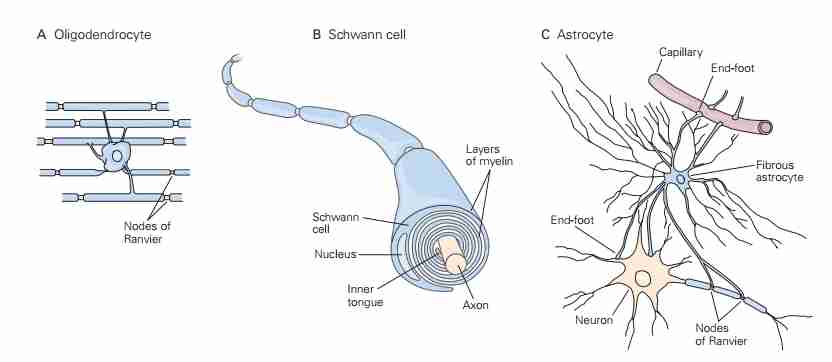
Astrocytes
Astrocytes are star-shaped glial cells predominantly found in the central nervous system (CNS) (brain and neural cord).
- Biochemical support for clearing things up and providing nutrients.
- They are involved in maintaining the blood-brain barrier, regulating ion and neurotransmitter concentrations in the extracellular environment,
- Providing metabolic support to neurons. Additionally, astrocytes contribute to synaptic plasticity by modulating neuro-transmission and facilitating the clearance of excess glutamate, preventing excitotoxicity.
Oligodendrocytes
Oligodendrocytes are specialized glial cells responsible for myelinating axons in the CNS. Unlike Schwann cells, which myelinate a single axon in the peripheral nervous system, a single oligodendrocyte can extend its processes to myelinate multiple axons. This myelin sheath enhances the speed of electrical conduction through saltatory conduction, which is critical for efficient neural communication. Oligodendrocytes also provide metabolic support to neurons and are implicated in neurodegenerative diseases such as multiple sclerosis, where their dysfunction leads to demyelination and impaired neural signaling.
Microglia
Microglia serve as the resident immune cells of the CNS, functioning as the first line of defense against pathogens and injury.
- They work as garbage removal cells. (toxic molecules removal).
- They constantly survey their environment and, upon detecting damage or infection, transition into an activated state to clear debris, remove apoptotic cells, and release inflammatory mediators. While microglia are essential for neuroprotection, their overactivation has been linked to chronic neuroinflammation, which plays a role in neurodegenerative diseases such as Alzheimer’s and Parkinson’s.
- Recent research suggests that microglia also contribute to synaptic pruning, a crucial process in neural circuit refinement during development.
They share many properties with macrophages found in other tissues, and are primarily scavenger cells that remove cellular debris from sites of injury or normal cell turnover.
Schwann Cells
Schwann cells are the principal myelinating glial cells in the peripheral nervous system (PNS). Unlike oligodendrocytes, each Schwann cell wraps around a single axon segment, forming the myelin sheath necessary for rapid signal transmission. Beyond myelination, Schwann cells support axonal regeneration following injury, a capability that is notably limited in the CNS. They achieve this by releasing growth factors, clearing myelin debris, and guiding regenerating axons toward their targets. Due to their regenerative potential, Schwann cells are being investigated for therapeutic applications in spinal cord injuries and peripheral nerve repair.